
A single diagnostic x-ray delivers a very small dose of radiation, and the added cancer risk from that one exam is too small to measure directly. Repeated high-dose scans, especially CT, add up and raise measurable risk over time. The EPA, FDA, and American Cancer Society agree: a chest x-ray delivers about 0.1 mSv, compared to roughly 3 mSv a year from natural background radiation.
TL;DR:
- Repeated high-dose scans like CTs contribute significantly to cumulative radiation exposure and increase measurable cancer risk.
- Low-dose diagnostic X-rays show no evidence of causing detectable cancer increases, with risk estimates based on conservative models that tend to overstate danger at typical doses.
- Children and pregnant women require additional precautions, including dose-optimized protocols and shielding, due to higher radiosensitivity and fetal vulnerability.
- Patients should inquire about the necessity, radiation dose, and alternative imaging methods, and keep track of prior scans to minimize unnecessary exposure.
Table of Contents
- How Much Radiation Is in an X-Ray, Really?
- How Does X-Ray Radiation Affect Cancer Risk?
- Who Needs Extra Caution With X-Ray Exposure?
- Does Getting Frequent X-Rays Add Up Over Time?
- What Should You Ask Before Getting an X-Ray?
- Why Effective Dose Numbers Are Estimates, Not Guarantees
- Can Too Much X-Ray Radiation Cause Symptoms?
- What Rules Govern X-Ray Safety in the United States?
- Our Perspective on Imaging Safety and Patient Conversations
- Get Answers About Your Own Imaging Needs
- Sources
- FAQ
How Much Radiation Is in an X-Ray, Really?
Numbers help more than reassurance alone. A chest x-ray delivers about 0.1 mSv, which the EPA equates to roughly 10 days of ordinary background exposure from soil, air travel, and cosmic rays. A dental bitewing sits even lower, at 0.004 to 0.005 mSv. Mammograms run between 0.13 and 0.36 mSv depending on breast density and the number of views taken.
CT scans are a different category entirely. A CT of the abdomen and pelvis averages 7.7 to 10 mSv, according to the American Cancer Society, and some interventional fluoroscopy procedures can reach tens of millisieverts depending on how long the procedure runs.
| Imaging test | Typical effective dose | Roughly equal to |
|---|---|---|
| Dental bitewing | 0.004–0.005 mSv | A few hours of background radiation |
| Chest x-ray | ~0.1 mSv | About 10 days of background radiation |
| Mammogram | 0.13–0.36 mSv | about 10 days of background radiation |
| Abdomen x-ray | 0.004 to 0.005 mSv | about 10 days of background radiation |
| CT chest or abdomen | 7.7–10 mSv | roughly 3 years of background radiation |
| PET/CT | Often around 10 mSv combined | roughly 3 years of background radiation |
These figures come from population averages, not your specific body. The Health Physics Society notes that equipment settings, patient size, and the specific protocol used all shift the actual dose up or down, sometimes substantially. Treat the table as a way to understand scale, not a personal prescription.
How Does X-Ray Radiation Affect Cancer Risk?
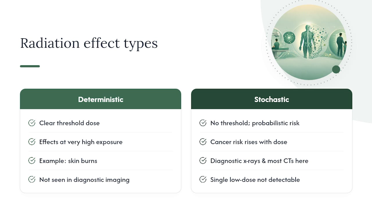
Radiation effects fall into two categories, and only one applies to routine imaging. Deterministic effects happen above a clear threshold dose, like skin burns from very high radiation exposure, and they simply don't occur at diagnostic imaging levels. Stochastic effects are probabilistic. The chance of a cancer developing rises with dose, but there's no threshold below which the risk is zero and no way to predict who, specifically, might be affected.
This distinction matters because it shapes how scientists talk about risk at low doses. Diagnostic x-rays and most CT scans fall in the stochastic category, and the American Cancer Society is direct about the limits of what we actually know: a single low-dose exam has never produced a detectable increase in cancer rates in epidemiological studies. The signal is too small to separate from background cancer rates that already run around 40% lifetime risk in the general population.
Risk estimates at these low doses come from extrapolation, mostly built on data from higher-dose exposures like atomic bomb survivor cohorts, then modeled downward using a linear no-threshold assumption. That's a scientifically conservative approach, not a measured fact, and it tends to overstate risk at diagnostic dose levels rather than understate it.
Clinical concern typically starts becoming concrete around cumulative doses near 100 mSv, a threshold cited by the American Cancer Society as the point where excess cancer risk becomes statistically detectable in large populations. A single x-ray or even a single CT scan sits well below that line. Repeated CT scans over months or years for a chronic condition are what push a patient's cumulative dose toward numbers worth tracking.
Who Needs Extra Caution With X-Ray Exposure?
Children are more radiosensitive than adults, mainly because their cells divide more rapidly and they have more remaining years of life for a stochastic effect to potentially develop. The CDC recommends dose-scaled pediatric protocols that reduce mA and kVp settings and tighten the imaging field, rather than using adult settings on a smaller body.
Pregnancy adds another layer of caution. Ultrasound is preferred whenever it can answer the clinical question, and when an x-ray is genuinely necessary, abdominal shielding and tight collimation keep fetal dose as low as possible. The FDA advises patients to tell their provider about a known or possible pregnancy before any imaging exam, every time, even for exams far from the abdomen.
Lifetime risk estimates also differ somewhat by sex and age at exposure, largely because younger patients have longer life expectancy remaining for any stochastic effect to manifest. Clinicians weigh this when deciding whether an exam's diagnostic value outweighs its theoretical risk for a particular patient.
Does Getting Frequent X-Rays Add Up Over Time?
There's no official "safe" cumulative dose limit for patients, unlike the annual limits set for radiation workers. That absence isn't an oversight. It reflects the fact that medical necessity, not a dose ceiling, is supposed to drive each imaging decision.
Cumulative dose still matters in practice. CT scans, interventional fluoroscopy procedures, and nuclear medicine studies contribute far more to a person's lifetime total than plain x-rays or dental imaging ever will. Someone managing a chronic condition with repeat CT monitoring can accumulate more radiation in a few years than most people see in a decade of occasional x-rays.
Keeping a personal imaging history, even an informal list of dates and exam types, gives your clinician something concrete to work from. If you're facing a third or fourth CT scan in a short window, it's reasonable to ask your doctor directly whether the cumulative total has been considered and whether a non-radiation alternative could answer the same question.
What Should You Ask Before Getting an X-Ray?
You have more influence over exposure than most people realize. A few direct questions can clarify whether an exam is worth it and whether it's being done as safely as possible:
- Why is this specific test needed, and how will the result change my treatment?
- What is the expected radiation dose for this exam?
- Are there non-radiation alternatives, like ultrasound or MRI, that could answer the same question?
- Will you use a pediatric or dose-optimized protocol if applicable?
- Can I get shielding for body parts outside the area being imaged?
Sharing prior imaging with a new provider avoids duplicate scans, which is one of the simplest ways to cut unnecessary exposure. If you've had a CT at another facility in the past year, mention it before agreeing to another one.
Pro Tip: Ask your imaging center whether their equipment uses automatic exposure control. Modern CT and fluoroscopy machines adjust output in real time based on body size, which can meaningfully lower dose compared to older fixed-setting equipment.
For complicated cases involving multiple prior scans, a radiology department's medical physicist can calculate a more precise cumulative dose estimate than the standard tables provide.
Why Effective Dose Numbers Are Estimates, Not Guarantees
The millisievert figures in dose tables represent effective dose, a calculation designed for comparing procedures and setting broad safety policy. It's not a direct measurement of the radiation your specific organs absorbed. RadiologyInfo explains that effective dose aggregates weighted risk across multiple organs into one number, which works well for population-level comparisons but can miss the mark for an individual patient or a highly localized procedure.
Body size, the exact protocol used, and the specific equipment all shift real exposure above or below the table average. A larger patient generally requires more radiation to produce a diagnostic-quality image than a smaller one on the same machine.
If you want a more personalized figure, tools like the RADAR Dose Risk Calculator extrapolate typical doses across a wide range of procedures. For genuine precision tied to your medical history, ask your imaging center directly. Many can pull the specific dose from your exam's metadata rather than relying on published averages.
Can Too Much X-Ray Radiation Cause Symptoms?
Radiation sickness from a diagnostic x-ray essentially doesn't happen. The doses involved sit thousands of times below the threshold where acute symptoms appear, and there is no documented case of standard diagnostic imaging causing radiation sickness in a patient.
Deterministic effects like skin reddening, hair loss at the exposure site, or cataracts have occurred, but only after unusually prolonged, high-dose exposure, typically from extended interventional fluoroscopy procedures lasting well over an hour, or from equipment malfunctions rather than routine exams. These effects require doses many times higher than any standard x-ray, CT, or even most fluoroscopy-guided procedures.
If they do occur, deterministic effects tend to show up at or near the exposed site rather than as a general body-wide illness. Skin changes might appear days to weeks after a prolonged procedure. Cataracts, when linked to radiation, typically develop years after chronic high-dose exposure, such as in occupational settings without adequate shielding, not from occasional diagnostic imaging.
What you should actually watch for after any medical procedure is far more mundane: unusual redness or irritation at a site where a prolonged interventional procedure was performed. That's worth mentioning to your doctor. Fatigue, nausea, or a general "off" feeling after a routine x-ray or standard CT scan has no established connection to radiation dose at those levels and is almost always explained by something else entirely, like contrast dye reactions, anxiety, or an unrelated illness.
What Rules Govern X-Ray Safety in the United States?
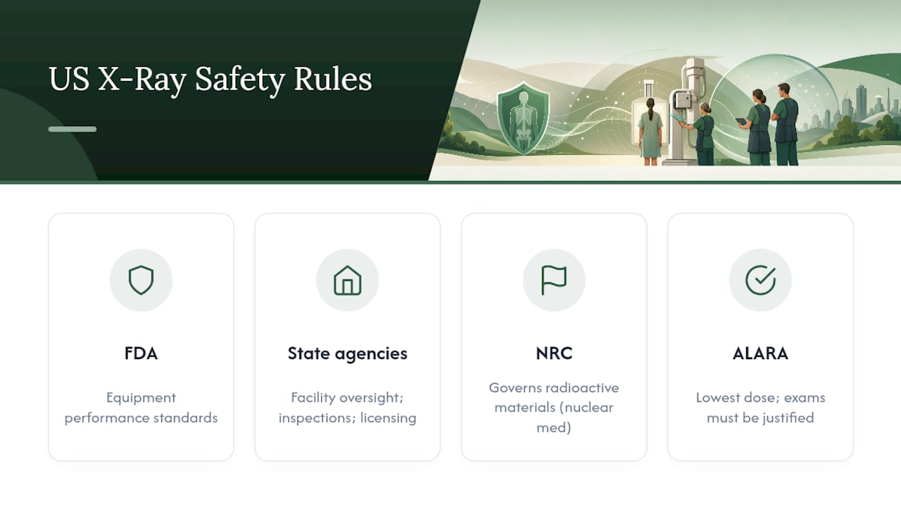
Medical imaging in the United States operates under layered regulation rather than a single rulebook. The FDA regulates the manufacturing standards for x-ray equipment itself, setting performance requirements that machines must meet before they reach a clinic or hospital.
State health departments handle facility-level oversight, including equipment inspections and technologist licensing, while the Nuclear Regulatory Commission governs radioactive materials used in nuclear medicine, a related but distinct category from standard x-ray and CT imaging.
The guiding clinical principle across all of it is ALARA, short for As Low As Reasonably Achievable. It's not a specific dose limit. It's a standard of practice requiring that every exam use the lowest radiation dose that still produces a diagnostically useful image, and that every exam be justified by genuine clinical need in the first place. The FDA and HPS both point to ALARA as the backbone of medical radiation safety, and internationally, the World Health Organization promotes similar dose-optimization and referral-criteria frameworks to reduce unnecessary imaging worldwide.
Population-level dose from medical imaging actually declined between 2006 and 2016 according to HPS data, driven largely by better equipment and more disciplined referral practices. That trend reflects a system that has gotten measurably more careful over the past two decades, not less.
Our Perspective on Imaging Safety and Patient Conversations
Garden State Medical Group applies ALARA in daily practice, using pediatric and low-dose protocols where they're clinically appropriate rather than defaulting to one-size-fits-all settings. We'd rather spend two extra minutes explaining why a scan is needed than have a patient walk out uncertain about it. Bring your prior imaging history to your next visit, and ask us directly about dose. That conversation should never feel like an inconvenience.
— Garden State Medical Group
Get Answers About Your Own Imaging Needs
Reading dose tables only goes so far. What actually matters is whether a specific test makes sense for your specific situation, and that's a conversation, not a chart. Garden State Medical Group's Radiology & Diagnostic Testing services are performed on-site, which means your provider can review results, discuss dose, and weigh alternatives with you directly instead of routing you through a separate imaging center with no context on your history.

If you're weighing whether a recommended scan is necessary, bring your prior imaging records to the visit, ask about pediatric or dose-optimized settings if the patient is a child, and request a dose estimate before the exam if you want one. You can also read more about when a radiology referral actually makes sense before your appointment. To schedule an imaging exam or a consult about a scan you've already had recommended, contact Garden State Medical Group directly through the radiology services page.
This article is general information, not a substitute for advice from a qualified doctor. Consult a qualified healthcare professional about your own circumstances before acting on anything here.
Sources
- Frequent Questions: Radiation in Medicine | EPA
- Understanding Radiation Risk from Imaging Tests | American Cancer Society
- Radiation Exposure From Medical Exams and Procedures | Health Physics Society (HPS)
- Radiation Dose from X-Ray and CT Exams | RadiologyInfo
- Questions and answers for physicians about medical x‑rays | FDA
FAQ
Can radiation from x-rays be harmful?
At the low doses used in routine diagnostic imaging, the cancer risk from a single exam is too small to measure directly. Risk becomes a legitimate concern mainly with repeated high-dose procedures like multiple CT scans over a short period.
How often can you safely get x-rays?
There's no official limit on the number of x-rays a person can have, since risk is tied to cumulative dose and medical necessity rather than a fixed count. Ask your clinician to track your imaging history if you're undergoing frequent scans, especially CT or fluoroscopy.
Which is safer, MRI or x-ray?
MRI uses magnetic fields and radio waves instead of ionizing radiation, so it carries no radiation-related cancer risk, unlike x-rays or CT scans. That said, x-rays remain the better and faster choice for many conditions, like detecting fractures, where MRI offers no diagnostic advantage.
Is one x-ray per year safe?
A single annual x-ray, such as a routine chest x-ray at roughly 0.1 mSv, adds a small fraction to your yearly background exposure of about 3 mSv and carries an extremely low individual cancer risk. Clinicians weigh that minimal risk against the diagnostic benefit every time a test is ordered.