
A normal carotid ultrasound means no significant narrowing and no urgent action needed. The findings that do call for prompt attention are severe stenosis (usually 70% or higher), a fully blocked artery, or plaque described as mobile or ulcerated. Everything else, including mild to moderate narrowing, typically leads to medical management and follow-up rather than an emergency call. What happens next depends heavily on whether you've had symptoms like a TIA or stroke.
TL;DR:
- Carotid artery occlusions, near-occlusions, mobile plaques, and ulcerations require immediate follow-up and often prompt further imaging like CTA or MRA for detailed assessment.
- A peak systolic velocity over 125 cm/s indicates at least 50% stenosis, while velocities above 230 cm/s suggest 70% or greater narrowing, influencing treatment urgency.
- Plaque stability, characterized as soft, ulcerated, or heterogeneous, increases stroke risk independently of stenosis degree and warrants expert evaluation.
- Most cases of mild to moderate stenosis with stable plaques are managed medically through lifestyle and medication, with invasive procedures reserved for severe cases or symptomatic patients.
- Follow-up imaging intervals typically range from six months to a year, especially if findings include moderate to severe stenosis or unstable plaque features.
Table of Contents
- Understanding Carotid Ultrasound Results: Reading Your Report
- The Numbers That Actually Matter: Stenosis, Velocities, and CIMT
- Why Plaque Appearance Matters as Much as the Percentage
- What Happens Next: Treatment Pathways Based on Your Results
- When Ultrasound Isn't Enough: Limits and Next Imaging Steps
- Getting Ready for Your Follow-Up Appointment
- Garden State Medical Group's Approach to Reading Your Results
- How Garden State Medical Group Supports You After Your Results
- Sources
- FAQ
Understanding Carotid Ultrasound Results: Reading Your Report
Your carotid ultrasound report is built in layers, and the fastest way to get your bearings is to skip to the bottom first. The Impression section (sometimes called "Conclusion") is where the radiologist summarizes what matters. Everything above it, technique notes, Doppler velocity tables, plaque descriptions, is the evidence that led to that conclusion.
Before you interpret anything, confirm which artery and which side each line refers to. Reports typically separate the right and left carotid systems, and within each side you'll see the common carotid artery (CCA), the internal carotid artery (ICA), and sometimes the external carotid artery (ECA). A finding in your right ICA does not tell you anything about your left CCA. Mixing them up is the single most common way patients misread their own results.
A few words carry more weight than the rest of the page:
- Occlusion means the artery is completely blocked.
- Near-occlusion describes a critically narrowed vessel with unusual flow patterns.
- Mobile plaque or ulceration suggests unstable material that could break loose.
- String sign refers to a trickle of flow through a severely narrowed segment.
If any of these appear in your impression, that's the part of the report your provider will act on first.
The Numbers That Actually Matter: Stenosis, Velocities, and CIMT
Carotid ultrasound reports lean heavily on numbers, and each one maps to a specific clinical category.
Percent stenosis is the headline figure. Under 50% is classified as mild narrowing. Between 50% and 69% is moderate. Severe stenosis covers 70% to 99%, and 100% means the artery is fully occluded. These categories drive nearly every downstream decision, from how often you get rechecked to whether a surgeon gets involved.
Doppler velocities are how the sonographer actually calculates that percentage. The internal carotid artery's peak systolic velocity (PSV) is the key measurement: a reading above roughly 125 cm/s suggests 50% or greater narrowing, and above about 230 cm/s points toward 70% or greater stenosis. Technicians also calculate an ICA/CCA ratio and look at end-diastolic velocity (EDV) to refine the estimate when the picture is ambiguous, which happens more often than you'd think in patients with irregular anatomy or calcified vessels.
By the numbers: ICA PSV over 125 cm/s flags 50%+ stenosis; over 230 cm/s flags 70%+ stenosis. CIMT under 0.9 mm is normal; over 1.0 mm is elevated; 1.5 mm or higher is often called plaque.
Carotid intima media thickness (CIMT) measures the wall of the artery itself, not a blockage. Most adults fall under 0.9 mm. A reading above 1.0 mm is generally considered elevated, a sign of early vessel aging even without a discrete blockage. When thickness reaches around 1.5 mm or more, it is typically classified as focal plaque rather than diffuse thickening.
Clinical guidelines commonly set the bar for intervention at 70% stenosis or higher in patients with symptoms, and 80% or higher in patients without symptoms, though your care team weighs your full health picture before recommending anything invasive.
Why Plaque Appearance Matters as Much as the Percentage
A mid-range stenosis number doesn't tell the whole story. Two patients with identical 40% narrowing can carry very different stroke risk depending on what that plaque looks like under ultrasound.
Radiologists describe plaque texture using terms like hypoechoic (dark, often lipid-rich), heterogeneous (mixed composition), calcified (hard and generally more stable), ulcerated (surface breakdown), and mobile (loose material moving with blood flow). Soft, irregular, or ulcerated plaque tends to be more prone to rupture and embolization than smooth, calcified plaque, even when the artery itself isn't severely narrowed. Research on vulnerable plaque shows these features can raise stroke risk independent of stenosis severity, which is why modern vascular labs increasingly describe plaque character alongside the percentage.
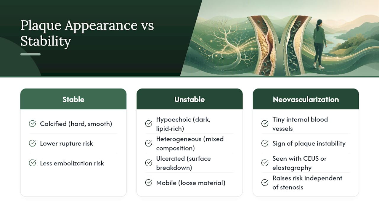
Some centers now use contrast-enhanced ultrasound (CEUS) or elastography to look for neovascularization, tiny new blood vessels growing inside the plaque that signal instability. These tools add useful detail but aren't universally available and carry their own limitations.
Pro Tip: Ask your provider directly whether your plaque was described as stable or unstable, and whether neovascularization was assessed. That single question often reveals more about your real risk than the percentage alone.
What Happens Next: Treatment Pathways Based on Your Results
Your report category generally sorts into one of a few care paths.
- Mild to moderate stenosis with stable plaque. Most patients here start with lifestyle changes, statin therapy, and blood pressure control. An antiplatelet medication like aspirin may be added depending on your broader cardiovascular risk.
- Moderate stenosis with unstable plaque features or new symptoms. This often triggers closer surveillance and a conversation about whether earlier intervention makes sense, even if the percentage alone wouldn't demand it.
- Severe stenosis (70% and above) or occlusion. This is where referral for carotid endarterectomy or stenting typically enters the conversation, especially if you've had a TIA or stroke tied to that artery.
Symptom status changes everything. A 60% stenosis in someone who's had a recent TIA is treated with more urgency than the same percentage in someone with no symptoms at all. Age, other health conditions, and surgical risk all factor into shared decisions about timing.
When Ultrasound Isn't Enough: Limits and Next Imaging Steps
Carotid ultrasound is operator-dependent, and calcium deposits or difficult anatomy can obscure the view. Near-occlusion can also distort velocity readings in ways that make the percentage estimate unreliable.
When your report includes certain language, expect a recommendation for additional imaging:
- Occlusion or near-occlusion findings
- Mobile plaque or ulceration
- String sign
- Technically limited or inconclusive study
CT angiography (CTA) or MR angiography (MRA) usually follows in these cases, since both map the artery's anatomy more precisely and help a surgeon plan intervention if one is needed.
Getting Ready for Your Follow-Up Appointment
A little preparation makes your follow-up visit far more productive.
- Bring the full report, images included if you can get them, not just the summary page.
- Write down any recent symptoms: dizziness, vision changes, weakness, or slurred speech, even brief ones.
- List every medication and supplement you're currently taking.
- Ask specifically about your percent stenosis, your plaque description, and whether either has changed from a prior scan.
While you wait for that appointment, focus on what you can control now: quitting smoking, keeping blood pressure and cholesterol in range, and taking prescribed medications consistently. Watch for sudden weakness, facial drooping, or trouble speaking, and treat any of those as an emergency, not a wait-and-see situation.
Pro Tip: If your report doesn't mention a recommended re-scan interval, ask for one directly. Surveillance timing varies by findings, sometimes six months, sometimes a year or more, and it shouldn't be left unclear.
Garden State Medical Group's Approach to Reading Your Results
A multidisciplinary approach involves pairing radiology with cardiopulmonary care so abnormal findings don't sit in isolation. When a report raises concerns, your radiology results connect directly to a follow-up visit and a coordinated care plan.
— Garden State Medical Group
How Garden State Medical Group Supports You After Your Results
Getting a carotid ultrasound report is one thing. Knowing what to do with it is another, and that's where coordinated care makes the difference. Garden State Medical Group offers on-site Radiology & Diagnostic Testing for follow-up imaging, paired with Cardiopulmonary Care for patients whose results point toward ongoing cardiovascular risk management.
If your report flagged moderate or severe stenosis, elevated CIMT, or plaque features worth watching, the next step is a visit where your provider reviews the images alongside your symptom history and risk factors, not just the percentage on the page. The Heart Smart Program builds ongoing monitoring and risk-factor control around exactly these kinds of findings. When you schedule, bring your full report and a list of current medications so your provider can move straight to the questions that matter. Call your nearest North Bergen or Secaucus location, or request an appointment through Primary Care to get your results reviewed and a follow-up plan in place.
Sources
- Circulation / AHA — carotid stenosis intervention thresholds
- Advances in noninvasive carotid wall imaging with ultrasound: narrative review (PMC)
This article is general information, not a substitute for advice from a qualified doctor. Consult a qualified healthcare professional about your own circumstances before acting on anything here.
FAQ
What Are Good Numbers on a Carotid Ultrasound?
A normal result shows less than 50% stenosis, ICA peak systolic velocity under roughly 125 cm/s, and CIMT under 0.9 mm. These numbers generally mean no significant blockage and no immediate treatment change.
What Are the Warning Signs of a Blocked Carotid Artery?
Sudden weakness or numbness on one side of the body, trouble speaking, vision loss, or facial drooping are classic warning signs and should prompt emergency care immediately. These symptoms can indicate a TIA or stroke tied directly to carotid narrowing.
What Percentage of Carotid Blockage Is Acceptable?
Intervention is commonly considered once narrowing reaches 70% in symptomatic patients or 80% in asymptomatic patients.
What Does an Abnormal Carotid Ultrasound Look Like?
The specific wording in the impression, especially terms like occlusion or near-occlusion, tells you how quickly follow-up should happen.
Can Garden State Medical Group Help Interpret My Report?
Yes. Garden State Medical Group's Radiology & Diagnostic Testing team can review your carotid ultrasound results alongside your primary care and cardiopulmonary providers to determine appropriate next steps.